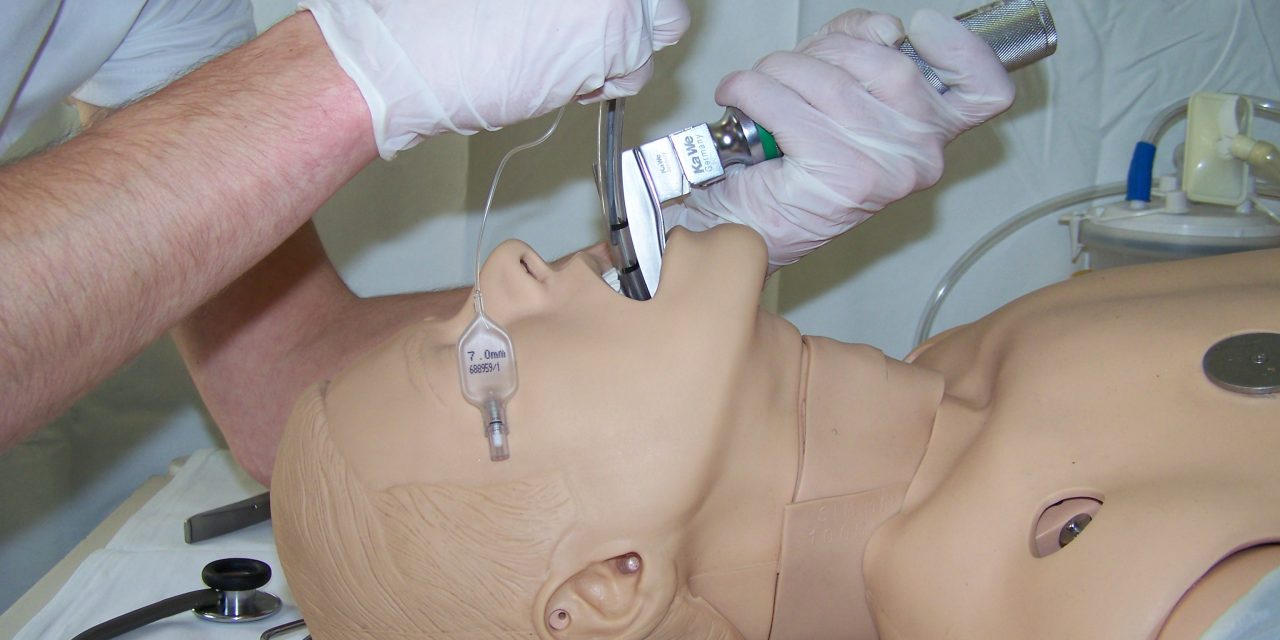
Endotracheal intubation is a procedure to manage and secure the airway by placing an endotracheal tube directly into the trachea. It is also the most reliable airway securing technique that completely secures the airway and enables optimal ventilation and oxygenation of the patient.
Personnel performing intubation must be familiar with all necessary equipment used in basic and advanced airway management and must be skilled in performing this procedure.
Endotracheal intubation should not last longer than thirty seconds. Steps for endotracheal intubation include:
- Preoxygenation of the patient with a high flow of oxygen.
- Positioning the patient for intubation (slightly bent head with a small pillow under the occipital part, the head is extended).
- Open the patient’s mouth, hold the laryngoscope in the left hand and insert it into the right side of the patient’s mouth, moving the tongue with the laryngoscope to the left until the thickened pharyngeal cartilage (uvula) becomes visible.
- Slowly move the laryngoscope over the base of the tongue until the epiglottis is visible.
- Place the tip of the laryngoscope blade in the valicle (between the base of the epiglottis and the base of the tongue).
- Lift the laryngoscope blade upwards along the line of the laryngoscope.
- This maneuver raises the epiglottis and exposes the vocal cords.
- Once the larynx is visible, on either side of the vocal cords, insert the endotracheal tube.
- The tube is usually inserted through the right side of the mouth and is usually inserted up to the 21 mark for men and 23 for women.
- If there is doubt about the position of the tube, remove it, re-oxygenate the patient and repeat the procedure.
- After inserting the tube, it is sufficient to inflate the cuff to prevent air leakage during inhalation and secure the airway.
- Connect the self-inflating balloon to the tubing, which is connected to high-flow oxygen.
- Listen to the lungs and observe the rise of the chest during inhalation.
- Listen in the epigastrium for the presence of gastric dilatation and along the median axillary line on both sides of the chest.
- Confirm the position using a capnograph/capnometer, which measures the amount of carbon dioxide exhaled.
- If the capnograph/capnometer detects carbon dioxide, it can be assumed that the tube position is correct.
- After confirming the correct position of the endotracheal tube, it should be secured.
- If only the right side of the chest rises during breathing, the cuff should be deflated, indicating that the tube is in the right bronchus, remove the tube by 1-2 cm and re-inflate the cuff. After that, check the position of the pipe again.
- Intubation is performed after the patient has been given muscle relaxation to achieve airway relaxation. The patient must be unconscious and must not have protective reflexes.
If the intubation is successful, after inflating the balloon on the endotracheal tube, confirming the position of the tube and securing the tube with the stabilizer, pressure on the laryngeal cartilage is no longer necessary.
If intubation is unsuccessful, the patient should be ventilated using a bag-valve-mask and 100% oxygen connected to a reservoir bag while maintaining pressure on the laryngeal cartilage.
In cases of anticipated difficult intubation, a video laryngoscope and equipment for difficult intubation can be used. Difficult intubation may include difficulty in visualizing the vocal cords or difficulty in tube placement due to distortion, swelling, or narrowing of the larynx and trachea.
After reoxygenation, an attempt will be made to re-intubate the patient, and if intubation fails after a re-attempt, an alternative airway (supraglottic device) or a surgical airway may need to be provided.
Confirmation of endotracheal tube placement can be objective or subjective, using pulse oximetry, wave capnography, and other methods. Undetected esophageal intubation and an improperly placed tube can be fatal to the patient, so all members of the emergency medical team must be aware of this. In addition to lung auscultation, the correct position of the endotracheal tube is best confirmed using a capnometer/capnograph to measure exhaled carbon dioxide.
A variety of intubation aids, supraglottic devices, and surgical airway management equipment may be used during difficult intubation. A bougie is an elastic endotracheal tube guide used when the vocal cords cannot be visualized. It is usually 60-70 centimeters long and is inserted blindly. The endotracheal tube is passed through the bougie.
Supraglottic devices are used when intubation by conventional means is unsuccessful. Various supraglottic devices are available, with I-gel and laryngeal mask being the most commonly used in Croatia due to their simplicity, non-invasiveness and economic profitability.


0 Comments