Although the name of this technique, the “Rapid Sequence Intubation Procedure,” suggests the rapid administration of drugs, the procedure itself is not rapid; it requires a certain amount of time.
We’ve broken down this advanced airway management technique into seven practical and essential stages:
- Preparation (for patient, equipment, team and drugs)
- Preoxidation (100% oxygen to the patient)
- Pretreatment (premedication if indicated)
- Paralysis with induction (sedation and relaxation)
- Protection (respiratory protection)
- Positioning (intubation procedure)
- Subsequent care after intubation (mechanical ventilation, maintenance of sedatives and muscle relaxants…)
Preparation of the patient, team, equipment and supplies for the procedure:
If the patient is conscious, the physician should explain the entire procedure to the patient, detailing the need for intubation due to their current condition. If the patient’s condition allows it, they should be placed in the optimal position for intubation.
The patient should be monitored, including ECG monitoring, oxygen saturation (SpO2), endotracheal CO2 (EtCO2) monitoring, and two large-bore intravenous lines should be established. Experience has shown that failure of this procedure is more common in the field, depending on successful establishment of intravenous access. Therefore, it is important to have two intravenous lines. In case of failure, an intraosseous approach should be considered.
Monitoring respiratory function is one of the most important indicators of a patient’s vital signs and includes two basic physiological functions: oxygenation and ventilation. Noninvasive methods for assessing these functions include noninvasive blood pressure (NIBP), measurement of arterial oxygen saturation (SpO2) using a pulse oximeter, and capnometry/capnography to measure endotracheal CO2 (EtCO2).
Monitoring can be performed in spontaneously breathing patients as well as in intubated patients.
Noninvasive measurement of arterial oxygen saturation using a pulse oximeter is the most widely used monitoring method and provides valuable information about the level of oxygen saturation. Pulse oximeters also measure heart rhythm. Normal values range between 95 and 100%, although accuracy can vary. Values may be inaccurate or limited in patients with severe hypoxemia, carbon monoxide poisoning, abnormal heart rhythms, hypoperfusion/hypotension, and when vasoconstrictive drugs are administered. Accuracy may also be compromised if the patient has painted nails, blood stains at the measurement site, strong ambient light, or if the oximeter is moved.
Capnography is a non-invasive method of monitoring carbon dioxide (CO2) levels in exhaled air (EtCO2) and can be administered via nasal cannulae or with the help of an adapter in intubated patients. Capnography uses infrared spectroscopy to continuously analyze the level of CO2 in respiratory gases, which is displayed as a wave on the monitor.
Normal values range from 35 to 45 mmHg. Values below 35 mmHg indicate hyperventilation, while values above 45 mmHg indicate hypoventilation. Capnography can warn of changes in metabolism and perfusion through elevated CO2 values.
Required equipment should include a defibrillator with ECG monitoring, 12-lead ECG, SpO2, and EtCO2 capabilities, a noninvasive blood pressure (NIBP) monitor, a mechanical ventilator capable of multiple modes of ventilation, and an aspiration system for airway management and suction of respiratory ways. Non-invasive blood pressure monitoring should be set to automatically measure at regular intervals. Early detection of hypotension can be facilitated by these measurements.
All equipment should be fully assembled and inspected upon handover by the previous emergency medical team, should be in good working order and regularly maintained by authorized technicians.
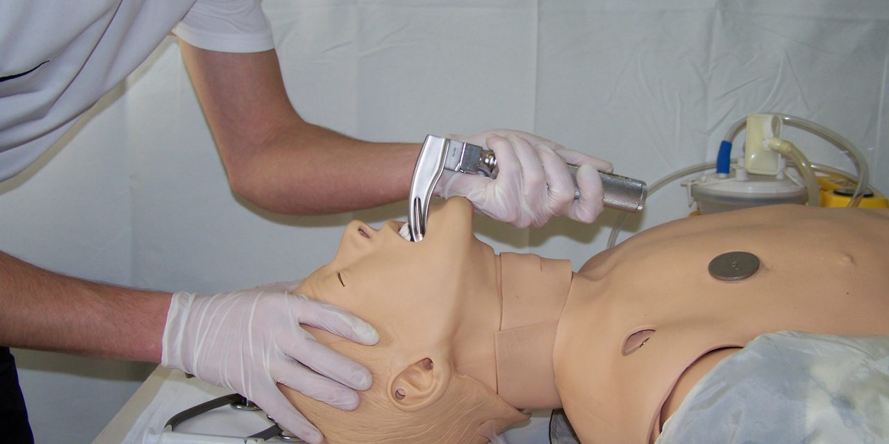
Respiratory equipment should be complete and ready for use. This includes equipment for basic and advanced airway management, such as laryngoscopes with replaceable batteries and paddles of various sizes with functional light, positive airway devices, and equipment for surgical airway access.
All drugs that will be used in the procedure should be prepared for administration before the intubation procedure, including dilution if necessary, and marked with special labels indicating the type and dose of the drug.
Members of the emergency medical team should be prepared for the procedure. Everyone should be equipped with appropriate personal protective equipment (gloves, goggles, protective gowns if necessary), protective clothing and footwear. Roles should be clearly defined based on formal education and additional professional training, and communication should be clear, loud and understandable.
Before the procedure, it is necessary to determine who will assist the doctor during intubation, who will apply pressure on the larynx cartilage, who will apply therapy, etc.


0 Comments